Figure 8 –
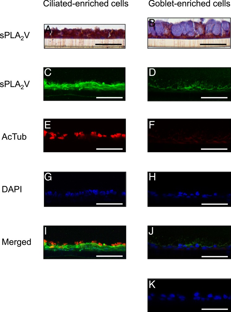
sPLA2 V is expressed in ciliated cells. sPLA2 V immunostaining and immunofluorescence with laser confocal microscopy of ciliated or goblet cell-enriched culture containing mucous granules grown for 14 d with PBS or with 5 ng/mL IL-13. A and B, Immunostaining. C-K, Immunofluorescence. A, C, E, G, I, PBS. B, D, F, H, J, K, IL-13. Immunofluorescence shows sPLA2 V (Alexa Fluor 488, green; C, D), acetylated α-tubulin (AcTub, Alexa Fluor 568, red; E, F), nuclei (DAPI, blue; G, H), and merged image of the double staining and DAPI (I, J). K, Negative control is HBE cells without the primary antibodies. Bar = 50 μm. See Figure 1 and 6 legends for expansion of abbreviations.

