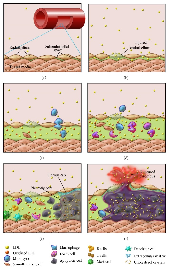
Figure 1.
A schematic drawing depicting the formation of atherosclerotic plaques. (a) In the wall of a normal artery, there is a very small subendothelial space in the intima between the endothelium and the smooth muscle cell layer in tunica media. (b) Hyperlipidemia and endothelial injury lead to the infiltration of LDL particles into the subendothelial space. (c) A large number of LDL particles are retained and subsequently oxidized in the subendothelial space, followed by monocyte infiltration (from lumen) and smooth muscle cell migration (from tunica media). (d) Monocytes and smooth muscle cells differentiate into macrophages, which engulf LDL and turn into foam cells, and are activated by oxidized LDL. SMCs are also activated, proliferate, and transform into lipid-laden foam cells. (e) Macrophage and smooth muscle foam cells undergo apoptosis; unbalanced apoptosis/efferocytosis results in necrotic core formation and unresolved inflammation. Other immune cell types also participate in the arterial wall inflammation. (f) Erosion of the fibrous cap caused by the matrix degrading enzymes secreted by the macrophages leads to unstable plaques, which eventually rupture and result in thrombus formation and adverse clinical events.

