
Figure 7. Apo-Lf but not holo-Lf reduced the cellular LIP.
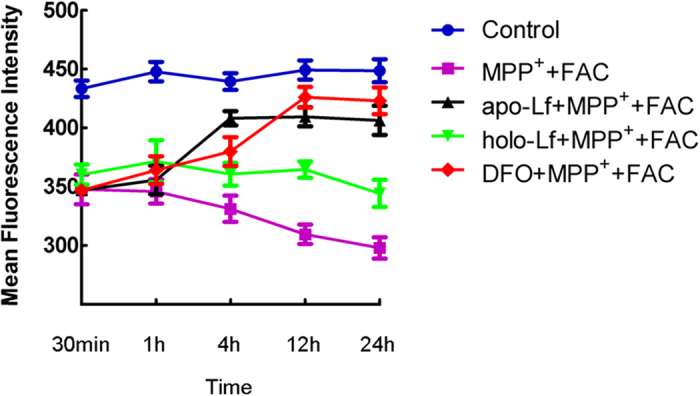
LIP in VM neurons was determined by the fluorescence intensity of calcein, an indicator of intracellular pool of free iron. The fluorescence intensity represented the mean value of VM neurons at each time point and was presented as the mean ± S.E.M. of six independent experiments. Results were carried out by two-way ANOVA followed by Student-Newman-Keuls test. There was a significant decrease in the fluorescence intensity in neurons treated with MPP++ FAC compared with the control, indicating increased free iron level. Fluorescence intensities increased in apo-Lf+ MPP++FAC or DFO+MPP++FAC groups, indicating decreased free iron levels in these cells. There was no obvious reverse in fluorescent intensity in holo-Lf+ MPP++FAC group, indicating holo-Lf did not change the cellular iron levels (two-way ANOVA, F = 39.835, P < 0.05, apo-Lf+MPP++FAC vs MPP++FAC; P < 0.05, DFO+MPP++FAC vs MPP++FAC; P < 0.05, holo-Lf+MPP++FAC vs apo-Lf+MPP++FAC and DFO+MPP++FAC;P > 0.05, holo-Lf+MPP++FAC vs MPP++FAC).