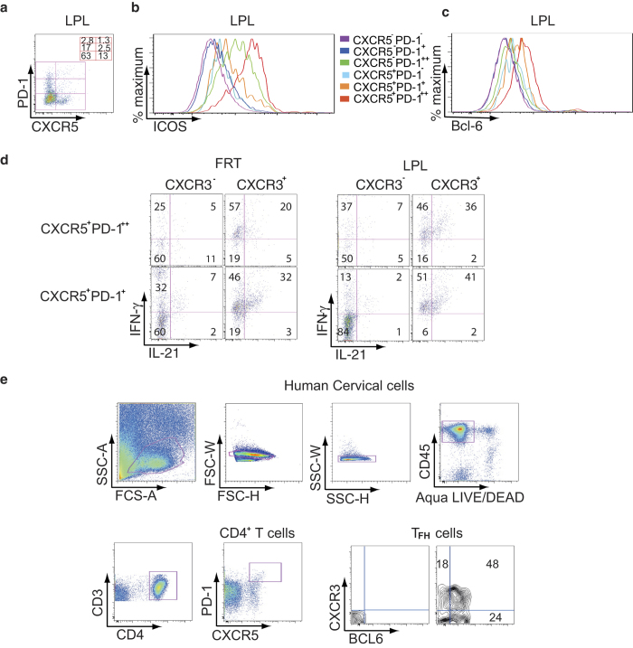
Figure 4. CXCR5+PD-1++ TFH cells in the gut and FRT of humanized DRAG mice express ICOS and BCL-6 and produce IL-21.
(a) A representative dot plot from two independent experiments of LPL depicts CD4+ T cells divided into 6 subsets based on CXCR5 and PD-1 expression. The frequency of CD4+ T subsets is shown. (b) and (c) Flow cytometry histograms from a representative experiment (n = 2) from LPL show ICOS (b) and BCL-6 expression (c) on CD4+ T cells. Cells were gated for hCD45 and Aqua LIVE/DEAD. Cells were then gated for CD3 and CD4. CD4+ T cells were gated for PD-1 and CXCR5 then for ICOS (b) and BCL-6 (c). (d) A representative dot plot from two independent experiments shows the cytokine production by TFH cells from FRT and LPL. Cells were stained for hCD45, Aqua LIVE/DEAD, CD3, CD19, CD8, IgD, CD38, and then sorted for CD4+ T cells and B cells. In a second sort, CD4+ T cells were stained for PD-1 and CXCR5 to sort for CXCR5+PD-1+ and CXCR5+PD-1++ CD4+ T cells. B cells were sorted for memory cells expressing CD38+IgD− cells. CXCR5+PD-1+ and CXCR5+PD-1++ CD4+ T cells were cultured with autologous memory B cells obtained from the same tissue. A schematic diagram of the sorting is shown in Supplementary Fig. 3. Stimulated FRT and LPL CD4+ T cells were stained for intracellular IL-21 and IFN-γ. (e) Endo- and ectocervix of human FRT were extracted from the muscular tissues and were digested with collagenase. The gating strategy is shown. Cells were gated for hCD45 and for Agua LIVE/DEAD followed by CD3 and CD4. CD4+ T cells were gated for PD-1 and CXCR5. CXCR5+PD-1++ TFH cells were unstained (left panel) or stained for CXCR3 and BCL-6 (right panel). A representative dot plot from two independent human cervical tissues is shown.

