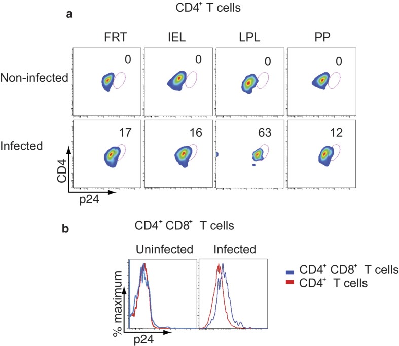
Figure 5. CD4+ T cells from FRT, IEL, LPL, and PP are highly permissive to HIV-1 infection.
Stimulated cells from the indicated tissues were infected with HIV-1 (primary US-1) for 2 days. (a) Flow cytometry shows intracellular staining of p24 in CD4+ T cells of the indicated tissues (upper panel: uninfected cells; lower panel: HIV-1 infected cells). (b) A representative overlay histogram shows the intracellular p24 staining in HIV-1 infected CD4+CD8+ T cells (blue line) compared to CD4+ T cells (red line) obtained from LPL (left panel: uninfected cells; right panel: HIV-1 infected cells). Cells were stained for both intracellular and extracellular CD4. A representative plot of two independent experiments is shown in (a) and (b).

