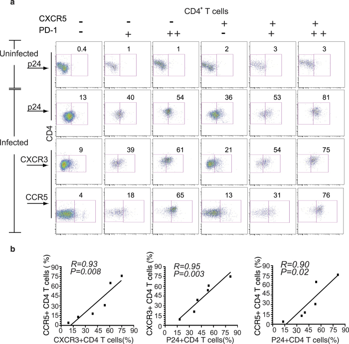
Figure 6. CXCR5+PD-1++ TFH cells are highly permissive to HIV-1 infection.
(a) LPL cells were stimulated with PHA for two days then left uninfected or infected with HIV-1 (primary US-1) for 3 days. HIV-1 infected CD4+ T cells were detected by intracellular staining with anti-p24 antibody. A representative flow cytometry dot plot showing p24 staining of non-infected CD4+ T cell subsets (top row); p24 staining of infected CD4+ T cell subsets (second row); CXCR3 positive CD4+ T cell subsets (third row) and CCR5 positive CD4+ T cell subsets (bottom row) are shown. Cells were stained for both intracellular and extracellular CD4. (b) Graphs show the correlation between the expression of CXCR3 and CCR5 (left), CXCR3 and intracellular p24 (middle) and CCR5 and intracellular p24 (right) CD4+ T cells from LPL. Linear regression, coefficient of correlation and P values are shown. Representative data from two independent experiments are shown.

