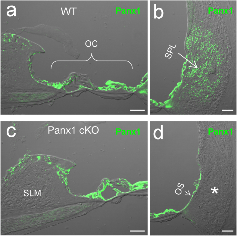
Figure 1. Panx1 deletion in the cochlea in Panx1 conditional knockout (cKO) mice.
a-b: Immunofluorescent staining for Panx1 in the WT mouse cochlea. c-d: Immunofluorescent staining for Panx1 in the Panx1 cKO mouse cochlea. An asterisk indicates that the area of type II fibrocytes in the cochlear lateral wall has no Panx1 labeling. OC: the organ of Corti, OS: outer sulcus cell, SLM: spiral limbus, SPL: spiral ligament. Scale bars: 25 μm.

