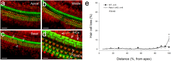
Figure 5. No substantial hair cell degeneration in Panx1 cKO mice.

a-c: The cochlear sensory epithelia of Panx1 cKO mice staining with phalloidin-Alexa Fluor-488 (green) and propidium iodide (PI, red). White arrows in panel c indicate scattered loss of outer hair cells (OHCs) in the basal turn. Scale bars: 50 μm. d: A high-magnification image. Inner hair cell’s (IHC) and OHC’s hair bundles are clearly visible. Scale bars: 20 μm. e: Hair cell loss accounting in Panx1 cKO and WT mice. Mice were P60-90 old. * P < 0.05, one-way ANOVA with a Bonferroni correction.
