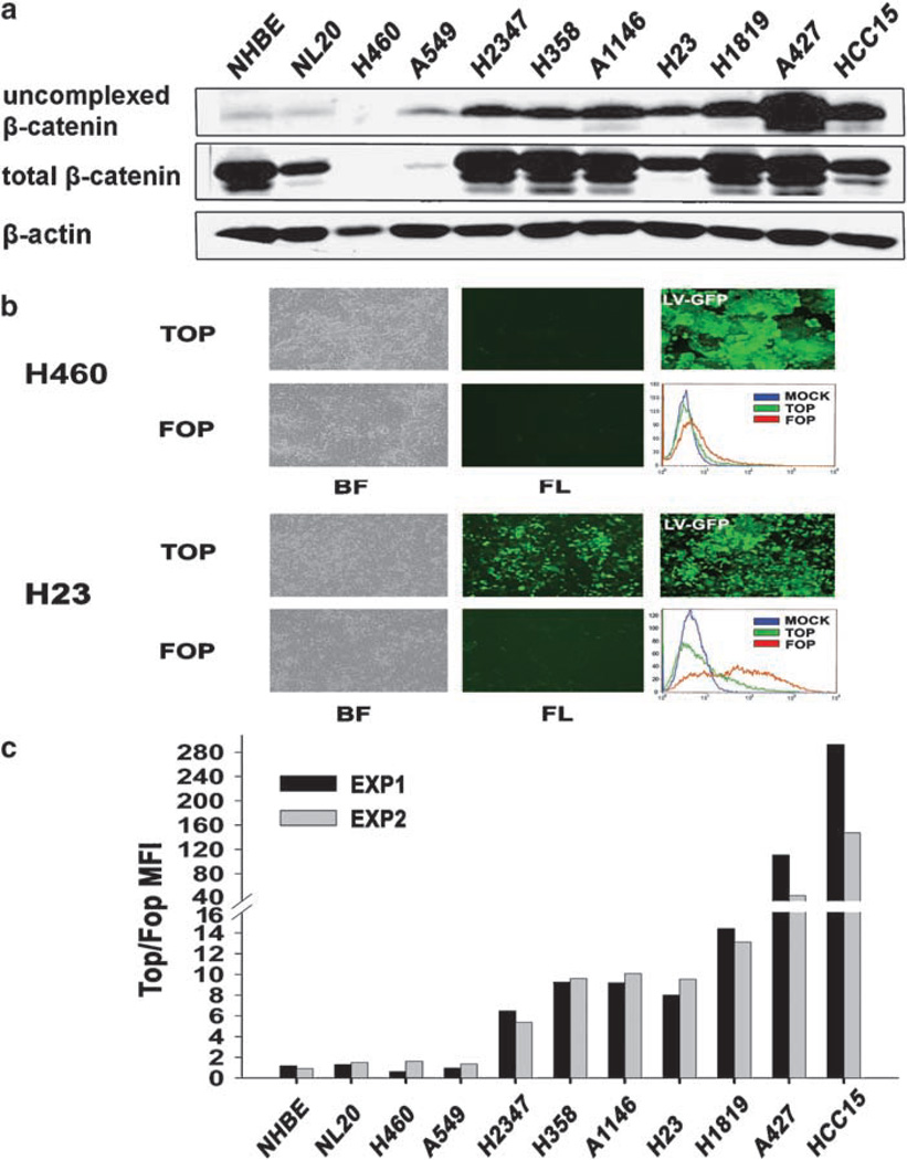
Figure 1.
Wnt signaling activation in human non-small-cell lung carcinoma (NSCLC) cell lines. (a) Total cell lysates (1 mg) were subjected to precipitation with a glutathione S-transferase (GST)-E cadherin fusion protein (Bafico et al., 1998). Total cell lysates (25 µg) and the GST-E-cadherin precipitates were subjected to immunoblot analysis with an mAb directed against β-catenin. (b) Fluorescence-activated cell sorting (FACS) analysis, phase contrast and fluorescence images of H460 (upper panel) and H23 (lower panel) NSCLC cells infected with TOP or FOP TCF-GFP reporter lentiviruses or with enhanced green fluorescent protein (EGFP) expressing lentivirus (LV-GFP). BF, bright field; FL, fluorescence. (c) Lentivirus-mediated TCF-GFP reporter activity in human NSCLC cells. Results are depicted as the ratio TOP/FOP GFP mean fluorescence intensity (MFI). Results from two independent experiments are shown.

