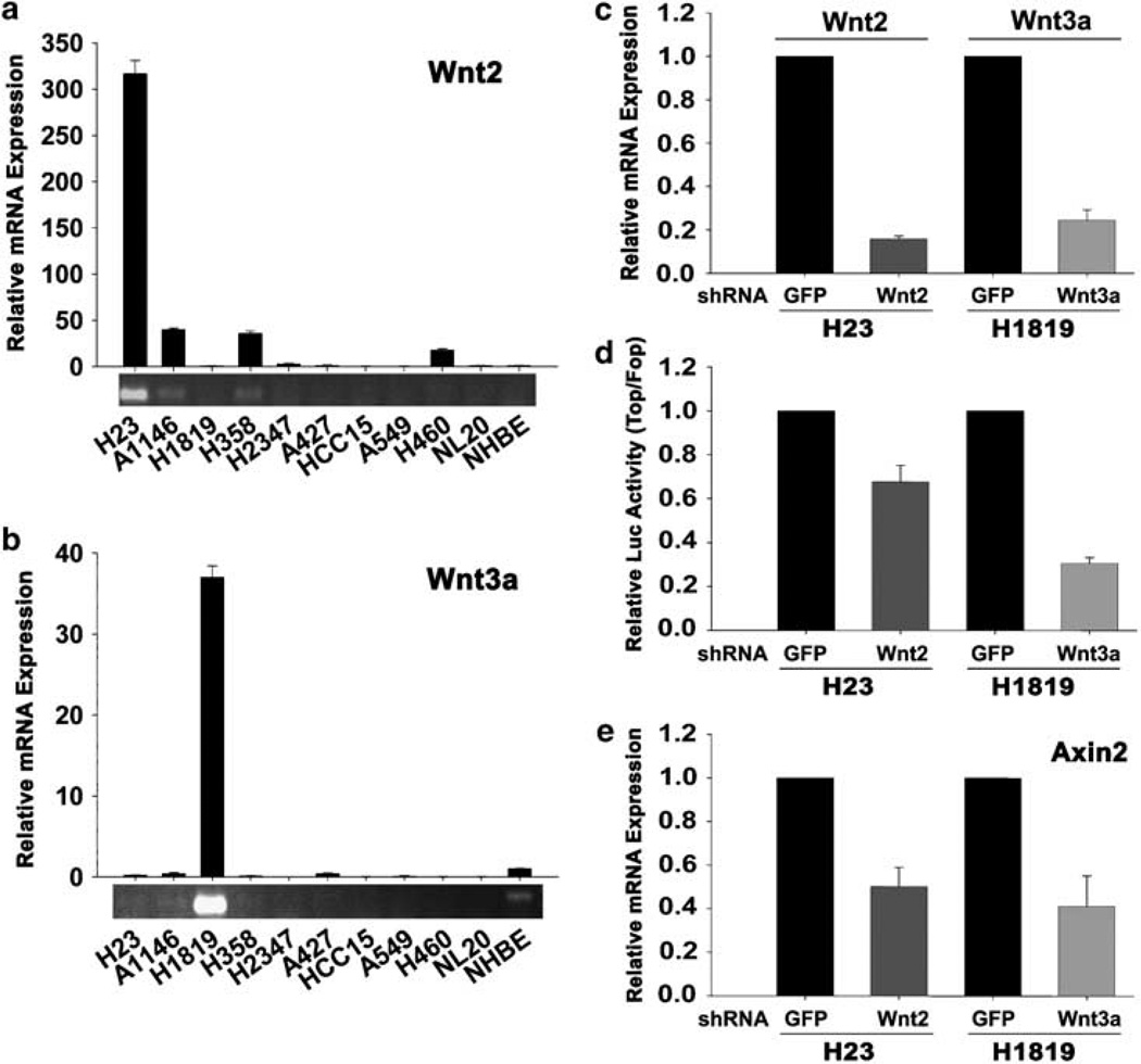
Figure 3.
Overexpression of Wnt2 and Wnt3a contributes to constitutive Wnt activation in autocrine non-small-cell lung carcinoma (NSCLC) cells. (a, b) Real-time PCR quantification of Wnt2 (a) and Wnt3a (b) expression in H23 and H1819 cells, respectively. To visualize relative expression levels of Wnt2 and Wnt3a, qPCR reactions were removed before saturation and PCR products were separated on 1.5% agarose gel and stained with ethidium bromide. (c) shRNA knockdown quantification of Wnt2 and Wnt3a. H23 and H1819 cells were infected with lentiviruses expressing shRNA targeting green fluorescent protein (GFP), Wnt2 or Wnt3a. (d) Effect of shRNA knockdown of Wnt2 and Wnt3a on TCF reporter activity. Luciferase reporter activity was calculated by dividing the TOP/RL ratio by FOP/RL ratio. Each column represents the mean ± s.d. of two independent experiments. (e) Effect of shRNA knockdown of Wnt2 and Wnt3a on axin2 mRNA expression. H23 and H1819 cells were infected with lentiviruses expressing shRNA targeting GFP, Wnt2 or Wnt3a.

