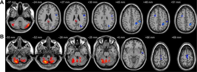
Figure 2.

Altered resting-state functional connectivity (rsFC) areas of the cerebellum posterior lobe (CPL) in the sleep deprivation group.
Notes: The rsFC areas of the left CPL were seen in the left precentral gyrus, left angular gyrus, left inferior frontal gyrus, left superior parietal lobule, left inferior parietal lobule, left postcentral gyrus, left precuneus, left posterior cingulate gyrus, right middle frontal gyrus, and bilaterally in the CPL (A), while the rsFC areas of the right CPL were seen in the left superior frontal gyrus, left middle frontal gyrus, left paracentral lobule, left superior temporal gyrus, left middle temporal gyrus, bilaterally in the cerebellum anterior lobe, and bilaterally in the CPL (B).
