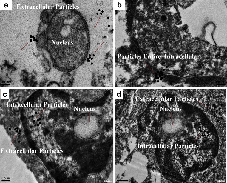
Fig. 9.
Transmission electron microscope images of MCF7 cells a after interaction with cisplatin-functionalized silica particles, inset shows extracellular particles, b the same sample suggesting endocytosis of the silica particles, inset shows particles at higher magnification, c and d control cells showing a smaller nucleus compared to the cisplatin-functionalized particle-treated cell

