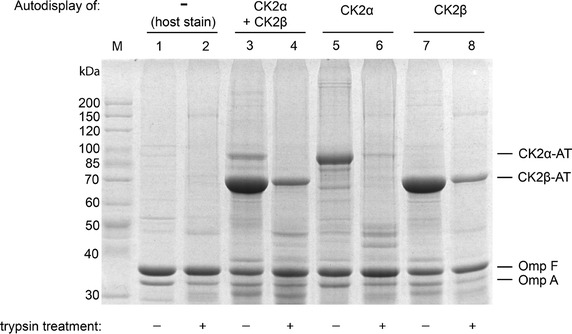
Figure 2.

Outer membrane preparation of CK2-presenting E. coli. Outer membrane samples were separated by SDS-PAGE (7.5% acrylamide) and stained with Coomassie brilliant blue. For autodisplay of the individual CK2 subunits, E. coli BL21(DE3) pCK2α (lanes 5 and 6) or E. coli BL21(DE3) pCK2β (lanes 7 and 8) was used, for autodisplay of both subunits in one strain E. coli BL21(DE3) pCK2α pCK2β (lanes 3 and 4) was used. A fraction of each whole cell sample was treated with trypsin before preparation of outer membranes to degrade surface presented proteins facing toward the extracellular medium (lanes 2, 4, 6 and 8). The host strain without any autodisplay construct served as a control (lane 1 and 2).
