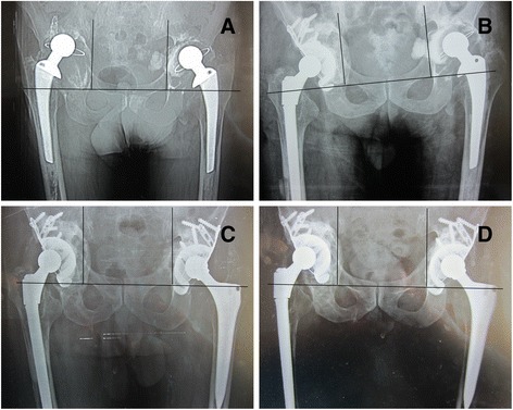
Fig. 1.

Preoperative, postoperative, and follow-up radiographic results. a Preoperative radiograph in a 65-year-old man shows massive acetabular bone defects on both sides. b Immediate postoperative radiograph shows reconstitution with a cage and morselized allograft in the right side. c After 3 months of right revision, the left side also completed the revision. Immediate postoperative radiograph shows reconstitution with a cage and a morselized allograft on the left side. d Radiograph taken at 7-year follow-up examination indicates that the bone grafts are incorporated relatively incompletely as we can see the sclerotic bone formed at the right side around the cage. And we also can see the acetabular cage move superiorly about 9 mm and more than a 5° change in the abduction angle on the right side, but the total reconstruction is still stable. This patient is asymptomatic, and his HSS is 98 at present
