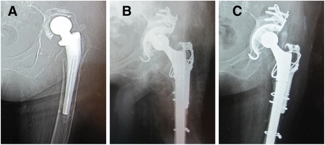
Fig. 3.

Preoperative, postoperative, and follow-up radiographs. a Preoperative radiograph of the hip of a 58-year-old woman shows massive acetabular bone defect. b Immediate postoperative radiograph shows reconstitution with a reconstruction cage and a massive morselized allograft. c Radiograph taken at 5-year follow-up examination indicates that the cup is stable and the bone graft is incorporated
