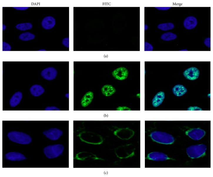
Figure 3.
Representative immunofluorescence staining pattern from a SLE serum with anti-MDM2 autoantibody positive (performed on Hep-2 antinuclear antigen tissue slides). (a) NHS was used as negative control; (b) a representative SLE serum with anti-MDM2 autoantibody positive demonstrated an intense nuclear staining pattern; (c) the same SLE serum used in panel (b) was preabsorbed with recombinant MDM2, and the nuclear fluorescent staining was significantly reduced.

