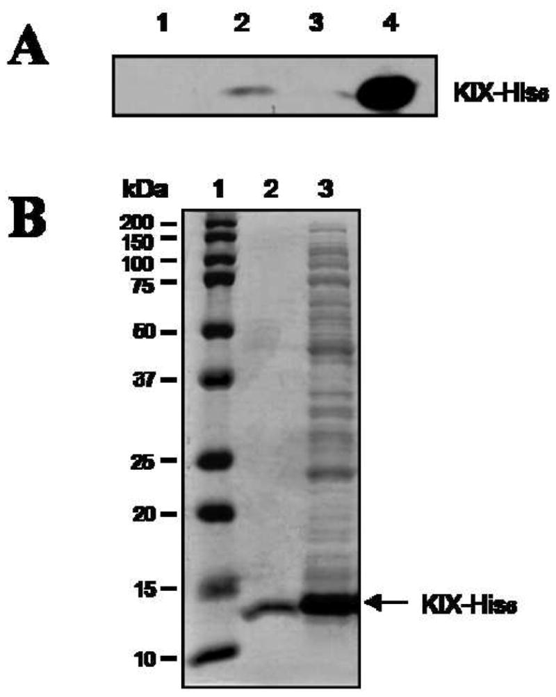
Figure 3.

Dimeric compound 3 retains His6-KIX protein specifically from a whole cell lysate. (A) Streptavidin beads were treated with DMSO (lane 1), 3-biotin (lane 2), and BHK2-biotin (lane 3) and then incubated with a cell lysate obtained from E. coli cells that overexpress His6-KIX. After washing, bound proteins were analyzed by Western blot with anti-His6 antibody. Lane 4: input. Note that this experiment was done under conditions of a large excess of KIX-His6 protein over immobilized peptoid and thus only a fraction if the input KIX domain could be retained by biotinylated 3. (B) Bound proteins were analyzed by Coomassie Blue staining. Lane 1: protein markers, lane 2: 3-biotin treated, lane 3: input (whole cell extract).
