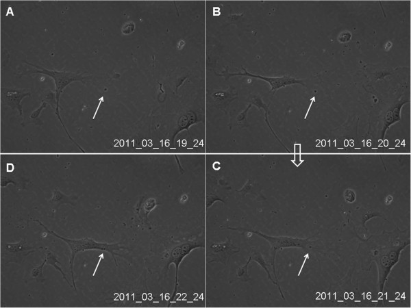
Figure 4.

Real-time dynamic observation of cultured lung telocytes through cell IQ cell culturing platform. The granular material (black arrow) appeared within the cellular frame, attracted cytoplasma, and was rounded with rich cytoplasma (A-D). Images were captured at 20 min intervals. Magnification is 200×. HPMECs were divided into two groups and cultured for 72 hours with DMEM and TCM respectively, and the proliferation of the cell was detected at 0 h, 12 h, 24 h, 48 h and 72 h. As showed in the figure, the proliferation of HPMECs cultured with TCM was faster than that of DMEM (P < 0.05).
