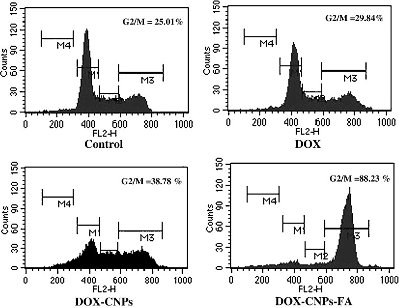
Fig. 6.
Cell cycle analysis by flow cytometry. For 48 h, 1 × 10⁵ cells/ml were treated with 5 μM of native DOX and equivalent concentrations of DOX in NPs (DOX-CNP and DOX-CNP-FA). Thereafter, to the treated cells, 20 μg/ml of propidium iodide solution was added and incubated at room temperature in dark for 30 min before flow cytometric analysis. Cell cycle distribution was determined by analyzing 15,000 ungated cells using a FACScan flow cytometer. Content of DNA is represented on the x-axis; number of cells counted is represented on the y-axis. Results are representative of one of three independent experiments

