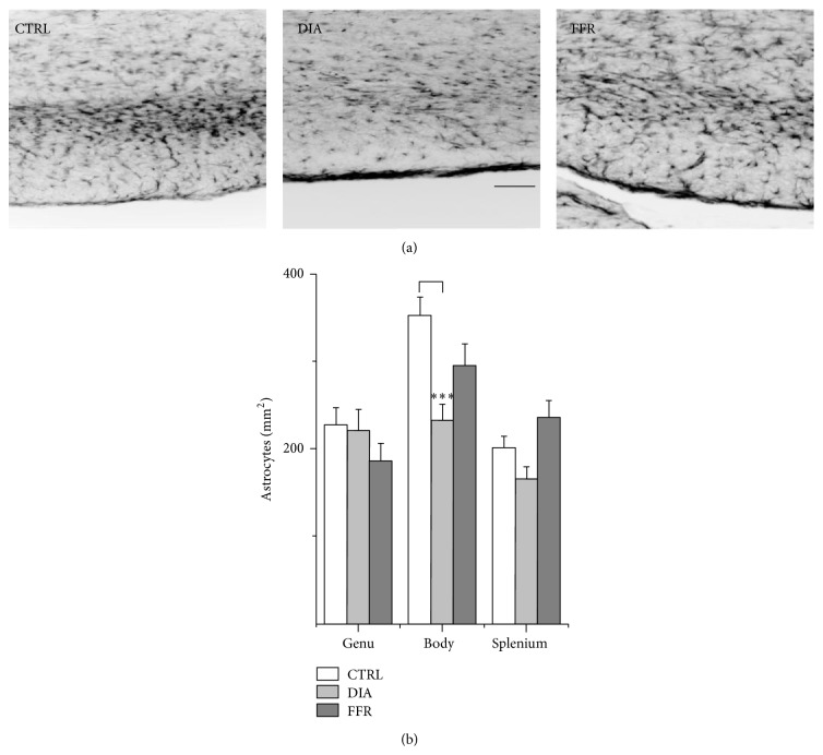
Figure 3.
Dehydration-induced anorexia (DIA) reduces astrocyte density in the body of rat CC. (a) Histological sections of the body of rat CC show GFAP staining in control (CTRL), dehydration-induced anorexia (DIA), and forced food-restricted (FFR) groups. (b) No differences in astrocyte density between control, DIA, or FFR groups were found in the genu (n ≥ 23) or the splenium (n ≥ 20). In contrast, a significant reduction in astrocyte density was observed in the caudal body (n ≥ 26) for the DIA group (−34%). Scale bar = 100 μm. Data are mean ± S.E.M. (∗∗∗ P < 0.001).

