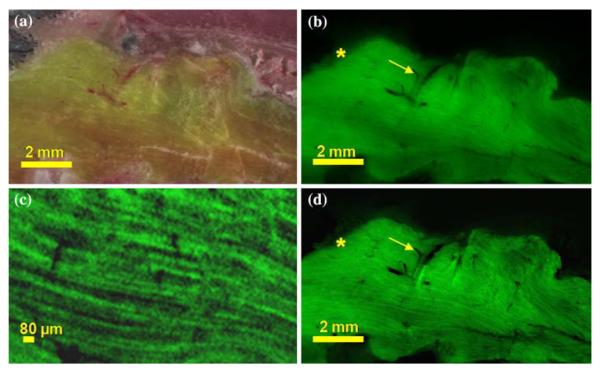
FIGURE 12.
Increased contrast in next-image processed cryo-images of GFP positive skeletal muscle. Brightfield and fluorescent cryo-images were collected of the GFP positive littermate in the parabiosis experiment. A brightfield cryo-image (a) of the GFP positive mouse skeletal is shown along with the corresponding fluorescent image (b). Out-of-plane fluorescence is visible along the periphery of the muscle (*) and inside of the vasculature (arrow). Processing removed out-of-plane fluorescent (c) resulting in significant improvement in image contrast as well as removing the halo effect (*) and out-of-plane fluorescence visible inside of the vasculature (arrow).

