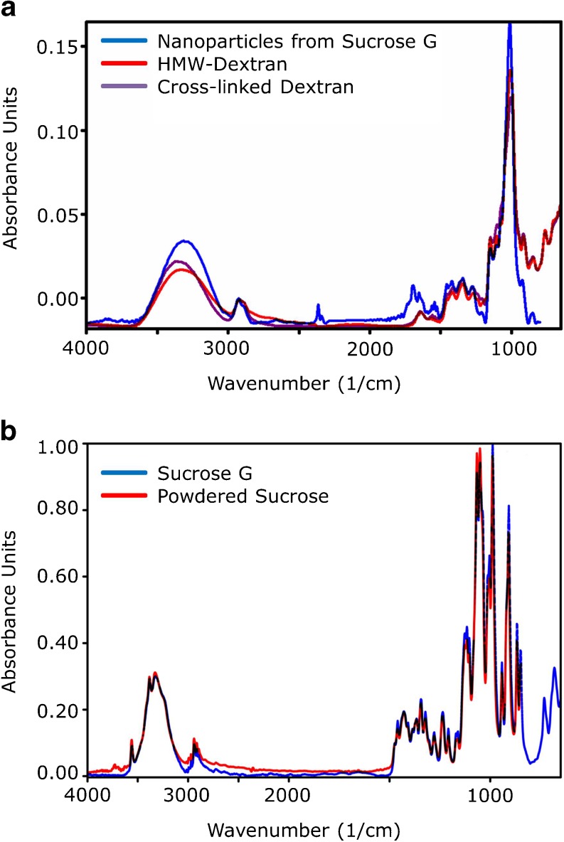
Fig. 5.
FTIR spectra recorded by FTIR microscopy overlaid with the best fitting entries of the S.T. Japan Europe GmbH database from 2009. (a) Recorded spectrum of vacuum dried nanoparticles isolated from sucrose G (blue) overlaid with the entries of high-molecular-weight (red) and cross-linked dextran (violet). (b) Recorded spectrum of unprocessed sucrose G (blue) overlaid with the entry of powdered sucrose (red).

