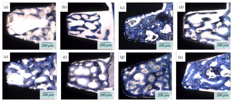
Figure 6.
Representative overview of the histological micrographs. Untreated calcium phosphate (CaP) implants at three (a) and six (e) weeks, respectively; untreated titanium (Ti) implant at three (b) and six (f) weeks, respectively; atmospheric pressure plasma- (APP-) treated CaP implants at three (c) and six (g) weeks; and APP-treated Ti group at three (d) and six (h) weeks.

