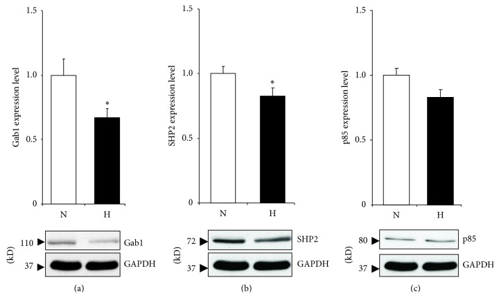
Figure 4.
Gab1 protein expression in rat cardiomyocytes following 24 h of hypoxic treatment. Cells were treated with hypoxic conditions (H) by incubating in a chamber with 5% CO2/0.2% O2 or normoxia (N) by leaving cells in normal CO2 incubator. After 24 h cells were lysed and western blot analysis performed using GAB1 (a), SHP2 (b), and p85 (c) antibodies. All results were normalized to GAPDH levels. Data are mean ± SEM. * P < 0.05 (n = 5).

