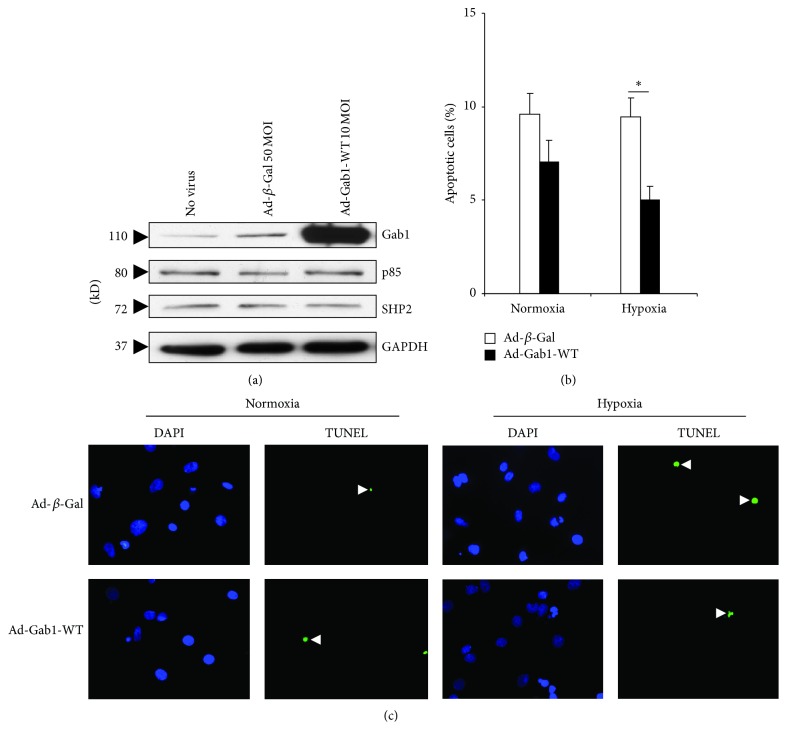
Figure 5.
(a) Protein expression level of Gab1, p85, and SHP2 in infected rat neonatal cardiomyocytes. Cardiac myocytes were infected with Ad-β-Gal (50 MOI) and Ad-Gab1-WT (10 MOI), for 24 hours. GAPDH was used as a loading control. (b) Apoptosis quantification of infected rat neonatal cardiomyocytes subjected to normoxia and hypoxia. Cardiac myocytes were infected with Ad-β-galactosidase (Ad-β-Gal) and Ad-Gab1-WT, for 24 hours. Data are mean ± SEM. * P < 0.05. (c) Representative pictures of the TUNEL assay performed on rat neonatal cardiomyocytes infected by Ad-β-Gal or Ad-Gab1-WT and subjected to normoxia or hypoxia. Arrows show apoptotic nuclei (green fluorescence). Cell nuclei were stained by DAPI (blue fluorescence). Magnification: ×400.

