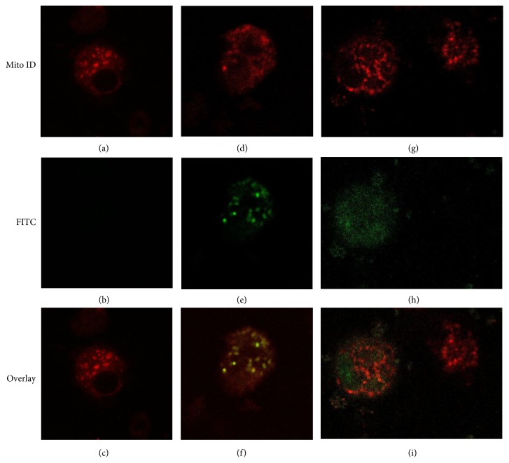
Figure 6.
Confocal microscopy of MDM-LPS cells after 24 hr incubation with 10 μM fullerene 1-FITC ((d), (e), and (f)) or fullerene 2-FITC ((g), (h), and (i)) or untreated control ((a), (b), and (c)), stained with Mito-ID (red fluorescence). Many fields were examined and over 95% of the cells displayed the patterns of the respective representative cells shown here.

