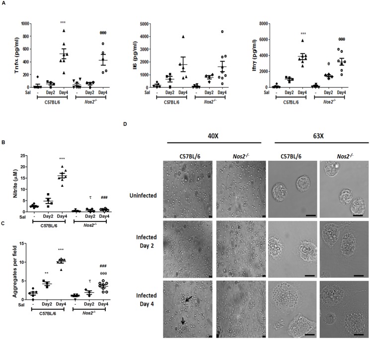
Fig 9. Nos2 mediates aggregation of APECs upon S. Typhimurium infection of mice.
The amounts of Tnfα, Il6 and Ifnγ (A) in the sera from uninfected and S. Typhimurium infected C57BL/6 and Nos2 -/- mice at day two and four post infection. The amounts of nitrite (B) and cell aggregates (C) of APECs isolated from uninfected and S. Typhimurium infected C57BL/6 and Nos2 -/- mice at indicated days post infection and cultured ex vivo for 24 h. Representative bright field microscopic images of APECs, either at 40X (Leica DMI6000B) or 63X (Leica TCS SP5), with the scale bar representing 20 μm, of APECs from uninfected or infected mice are shown (D). The data is representative of at least four independent experiments with a minimum of three mice per condition. Significance with respect to untreated C57BL/6 controls, untreated Nos2 -/- controls, S. Typhimurium infected C57BL/6 mice day 2 control and S. Typhimurium infected C57BL/6 mice day 4 control are represented as *, θ, τ, and # respectively.

