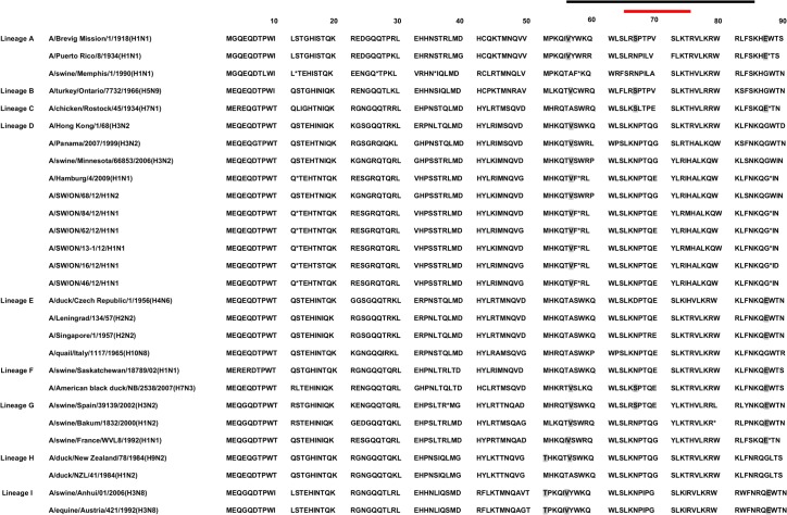
Fig 7. Presentation of polymerase B1-F2 (PB1-F2) variants of mammalian and avian influenza A viruses.
Nine PB1 lineages (A to I) have been described and presented. Colored lines at the top represent amino acids of the predicted helical region (black) and the putative mitochondrial targeting sequence (red). Amino acids that are considered to enhance viral pathogenesis are marked in gray (T51, V56, N66S, E87). The first stop codon has been shown by asterisk and following stop codons are indicated by asterisk.

