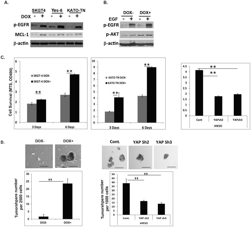
Figure 4. YAP activates EGFR signaling and mediates cell survival and CSCs properties.
A. Phospho-EGFR and MCL-1 were detected by immunoblotting in SKGT-4, YES-6 and KATO-TN cells transduced with YAP1S127 cDNA (PIN20YAP1) with (DOX+) or without (DOX-) doxycycline induction; B. Phospho-EGFR and phosphor-AKT were detected by immunoblotting in SKGT-4 cells transduced with YAP1S127 cDNA (PIN20YAP1) with (DOX+) or without (DOX-) doxycycline induction and treated with EGF at 50ng/ml. C. Cell growth of SKGT-4 (PIN20YAP) and KATO-TN (PIN20YAP) with (DOX+) or without (DOX-) YAP induction were determined using MTS as described in Materials & Methods to determine the rate of proliferation at three and six days. **P<0.05 (left and middle). Cell growth of JHESO control and YAP knockdown cells (YAP sh2 and sh3) were determined using MTS to determine the rate of proliferation at three days. **P<0.05 (right panel). D. Representative images of spheres in KATO-TN (PIN20YAP) cells with (DOX+) or without YAP1 induction (DOX-) (top); Representative bar graph demonstrating the sphere numbers in KATO-TN (PIN20YAP) cells with (DOX+) or without YAP1 induction (DOX-) (low) (left panel). Representative images of spheres in JHESO cells with control and its knockdown (YAPsh) cells (top); Representative bar graph demonstrating the sphere numbers in JHESO cells with control and its knockdown (YAPsh) cells (low) (right panel). Data are represented as mean and SD from three experiments. ***p<0.001.

