
Figure 6. Structural basis for negative cooperativity in ligand binding to Drosophila. EGFR.
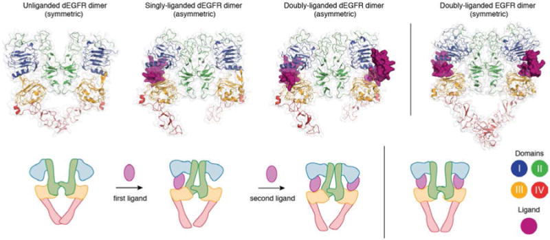
The unliganded (PDB ID 3I2T), singly-liganded (PDB ID 3LTG) and doubly-liganded (PDB ID 3LTF) dEGFR dimer. The structure of the doubly liganded human EGFR dimer (PDB ID 3NJP) is shown on the right.