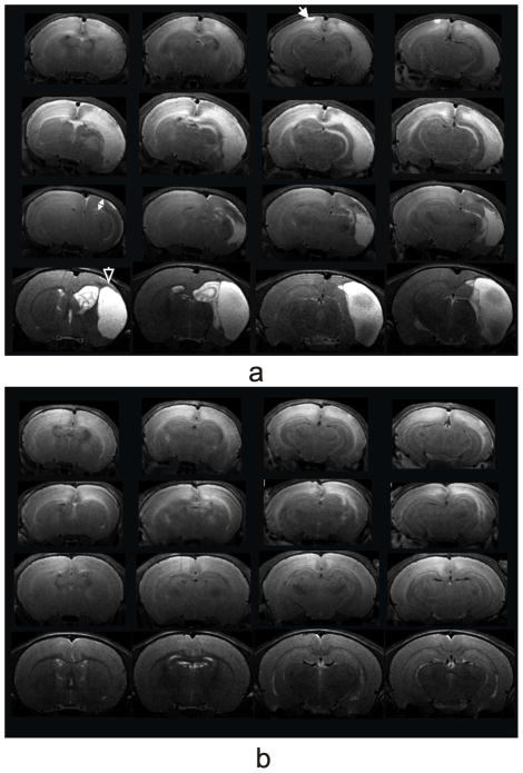
Fig. 2.
T2-weighted MRI coronal views from a male HI (a) and a male HI+ALCAR (b) rat on 24 h (top row), 72 h (second row), 7 d (third row), and 28 d (bottom row) post HI injury. The white closed arrow shows a lesion extended to the surface of the contralateral cortex in the HI rat. The white two-head arrow indicates a cortical thinning. The white open arrow shows a cyst with discrete boundaries in the HI rat.

