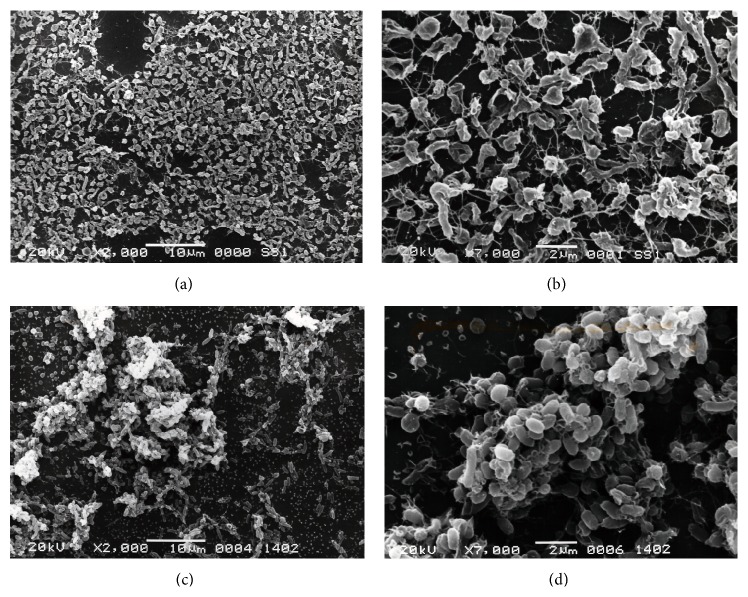
Figure 2.
SEM images of H. pylori strains SS1 ((a) and (b)) and TK1402 ((c) and (d)) biofilms. The 3-day biofilm of each strain on cover glass was investigated using SEM. Photographs were taken at low (×2000; (a) and (c)) or high (×7000; (b) and (d)) magnification. Scale bar (2 μm) is shown at the bottom of each electron micrograph.

