
Figure 3.
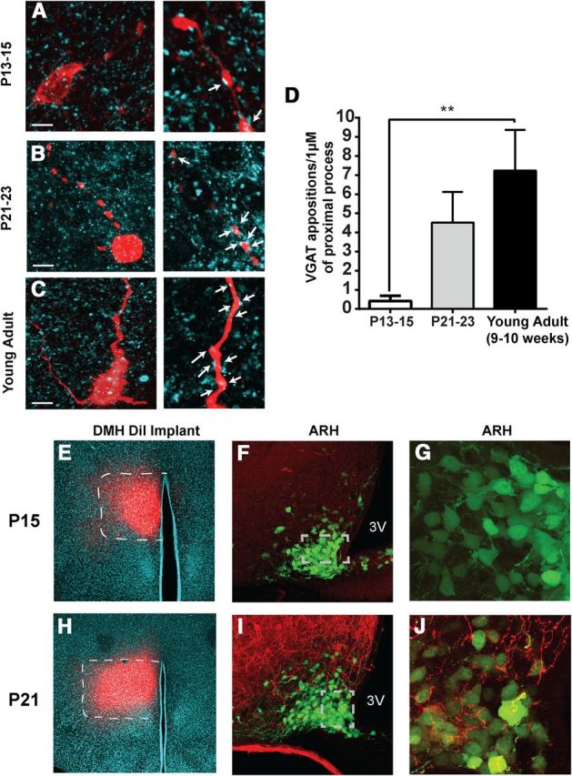
Age-associated changes in juxtaposed GABAergic terminals on NAG neurons and formation of projection pathways from the DMH to the ARH. A–C, Representative confocal images of NPY-GFP+ somas and proximal process filled with biocytin (red) during electrophysiological recording and VGAT (cyan) immunoreactivity. Left, Maximum projection images. Right, Zoomed 1 μm single optical slices in P13–P15 (A), P21–P23 (B), and young-adult mice (C). Arrows indicate juxtapositions (colocalization) suggesting possible synaptic contacts. Scale bar, 10 μm. D, Quantitative comparison of the number of VGAT synaptic boutons in close contact with biocytin-filled NAG proximal process (n = 2–3 optical sections per age, 31 animals). Results are shown as mean ± SEM; **p < 0.01, by ANOVA, post hoc Tukey's test. E–J, Representative confocal images of DiI implants (red) and DiI-labeled fibers (red) in the DMH and ARH during the third week of postnatal development. E, H, Appearance and distribution of a DiI-implant (red) in the DMH of postnatal mice at P15 and P21; DAPI staining (cyan), 10× (white dashed lines indicate the borders of the DMH). Confocal images of the ARH taken at 40× (F, I) and 63× with a digital zoom of two (G, J), showing the distribution of DiI-labeled (red) fibers and NPY-GFP+ neurons (green) in two mice (P15 and P21). Gray dashed lines define the limits of the high-magnification (63×) images. 3V, Third ventricle.