Figure 10.
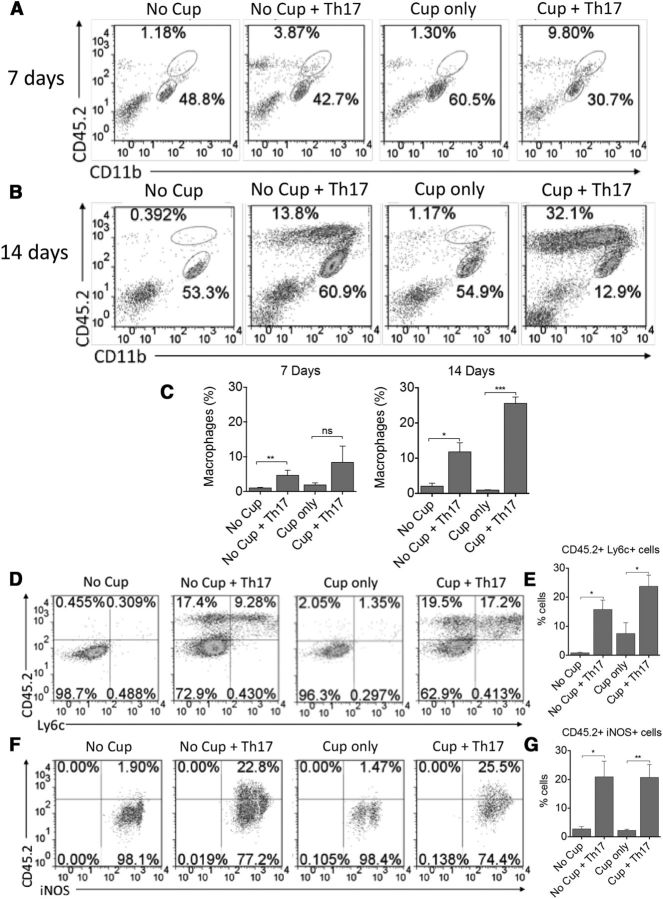
Peripheral inflammatory monocyte infiltration is increased in the CNS of cuprizone-fed animals at 14 d after transfer of myelin-reactive Th17 cells. Mononuclear cells were isolated from the brains of recipient animals at 7 d (A) and 14 d (B) after transfer of Th17 cells. The frequencies of CD11b+CD45hi monocytes and CD11b+CD45intermediate microglia were determined by flow cytometric analysis. Representative FACS plots are shown. Bar graphs represent quantitative analysis from 4 animals per group at 7 d (C) and 14 d (D) after transfer. The phenotype of CD11b+CD45hi monocytes was examined for the inflammatory markers Ly6C and iNOS (D, F). Representative FACS plots are shown following gating on CD11b+ cells. Bar graphs represent quantitative analysis from 6 animals per group at 14 d after transfer for CD45+Ly6C+ cells (E) and CD45+iNOS+ cells (G). All data are representative of three independent experiments. *p < 0.05 (two-tailed Student's t test). **p < 0.005 (two-tailed Student's t test). ***p < 0.001 (two-tailed Student's t test). ns, Not significant.

