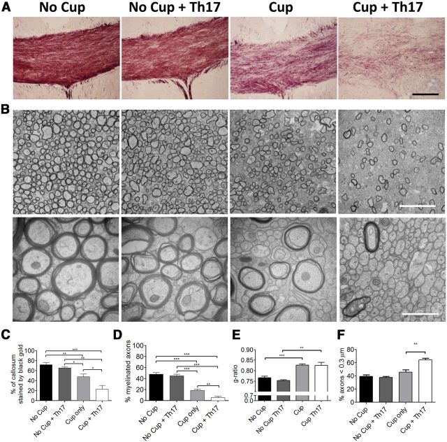
Figure 5.
Th17-transferred cells suppress endogenous remyelination after acute cuprizone exposure. Mice were fed 0.2% cuprizone for 4 weeks before transfer of Th17 cells and return to a regular diet. Remyelination and axonal integrity were evaluated 14 d after Th17 transfer. A, Black Gold staining demonstrates reduced myelin levels following transfer of Th17 cells to cuprizone-demyelinated mice. Scale bar, 100 μm. B, Electron microscopy of corpus callosum under indicated conditions. Left, original magnification ×10,000. Scale bar, 5 μm. Right, original magnification ×50,000. Scale bar, 1 μm. C, The percentage of the callosum that was stained by Black Gold was determined by dividing the area stained with Black Gold by the total area of the callosum. N = 5 or 6 animals per group. D, The percentage of myelinated axons was determined by counting myelinated axons and total axons in 10 images at ×10,000 magnification from 4 to 6 animals per experimental group. E, G-ratios are indicative of remyelination following cuprizone treatment. F, Small-caliber axons were counted at ×50,000. Th17 cells increase the abundance of small-caliber axons after cuprizone treatment. Data are mean ± SEM. *p < 0.05 (two-tailed Student's t test). **p < 0.005 (two-tailed Student's t test). ***p < 0.001 (two-tailed Student's t test). Data are representative of two independent experiments.

