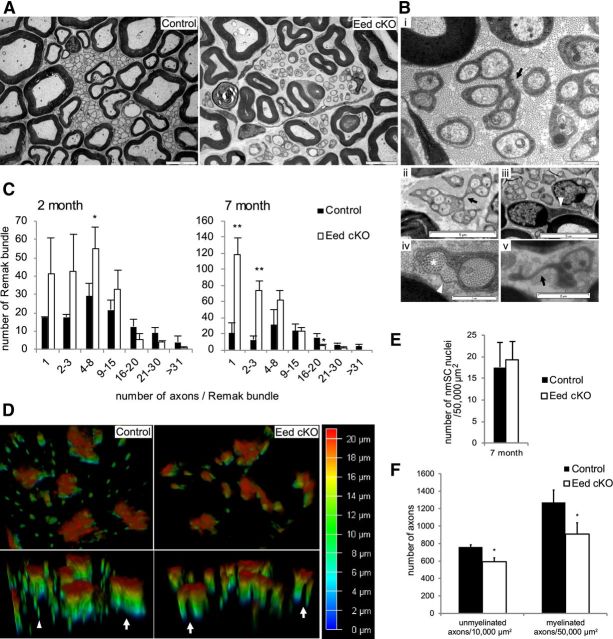
Figure 3.
Eed cKO nerves show Remak bundle fragmentation and the morphology change of nmSCs. A, EMs show fragmentation of Remak bundles in 7 month Eed cKO sciatic nerves compared with control nerves. Scale bars, 5 μm. B, EM of 7 month Eed cKO sciatic nerve. Arrows in i and ii point to where nmSC processes separate out to form two branches. An arrowhead in iii points to a single long process reaching a distant axon from a cell body. An asterisk in iv points to a collagen pocket, which is collagen surrounded by a nmSC process and a basal lamina (arrowhead). An arrow in v points to a free nmSC process. Scale bars: i, iv, 1 μm; v, 2 μm; ii, iii, 5 μm. C, The numbers of Remak bundles that are associated with >1000 randomly selected axons in control and Eed cKO sciatic nerves were grouped into seven categories based on the number of bundled axons. D, Transverse cryosections of sciatic nerves were stained with GFAP antibody and imaged using 3D deconvolution (color code indicates depth in the z-axis). The processes of Eed cKO nmSCs are separated while those of control nmSCs are in a bundle (arrows). An arrowhead indicates nonspecific staining. E, nmSC nuclei were counted in randomly selected fields that accounted for >45% of an entire sciatic nerve cross section from each animal and normalized per surface area (50,000 μm2). F, Unmyelinated and myelinated axons were counted in randomly selected fields that accounted for >8 and 44% of an entire sciatic nerve cross section from each animal and normalized per surface area (10,000 and 50,000 μm2), respectively. Fields that contain <15 unmyelinated axons were omitted for this quantification. Data: mean ± SD; **p < 0.005, *p < 0.05; n = 3 per genotype.

