
Figure 5.
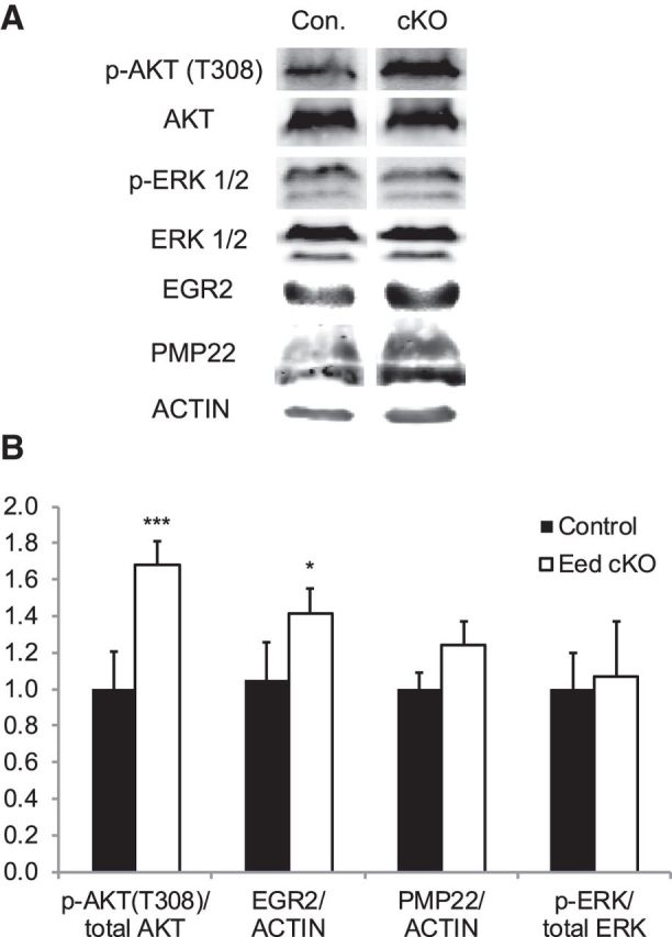
Increased activation of Akt in Eed cKO nerves. Western blot analysis of lysates from control and Eed cKO sciatic nerves using indicated antibodies. A, Blots of 2 month nerves for p-AKT/AKT and blots of 4 month nerves for p-ERK/ERK, EGR2/actin, and PMP22/actin. Lanes of control and Eed cKO samples were taken from the same image of the same membrane. B, Quantification of Western blot. p-Akt/Akt; n = 7 per genotype (1 month, n = 3 per genotype; 2 month, n = 4 per genotype). p-ERK/ERK, EGR2/actin, and PMP22/actin; 4 month, n = 4 per genotype. Data: mean ± SD; ***p < 0.00001, *p < 0.05.