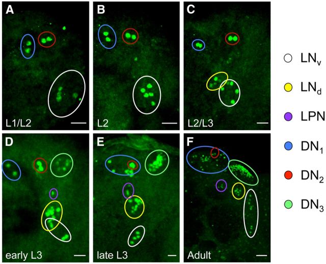
Figure 4.
Expression of CLK-GFP in the brain during larval development. Clk-GFP; Clkout flies were grown to the indicated stage (see Materials and Methods) and collected at ZT22. CNSs from these larvae were dissected, immunostained with GFP antibody, and imaged by confocal microscopy. A–F, Images of 24 h L1–L2 (A), 36 h L2 (B), 48 h L2–L3 (C), 60 h early L3 (D), >72 h late L3 (E), and adult (F) brains. For these developmental stages, 42 μm (L1–L2), 46 μm (L2), 46 μm (L2–L3), 44 μm (early L3), 50 μm (late L3) or 78 μm (adult) projected Z-series images are shown. Scale bars: A–E, 20 μm; F, 10 μm. The colored circles denote pacemaker neuron subgroups according to the key on the right. The different groups of pacemaker neurons were distinguished as described in Materials and Methods. All images are representative of 12 or more brain hemispheres.

