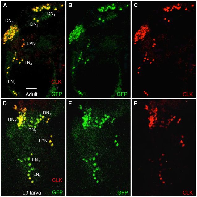
Figure 9.
GFP-CYC expression is restricted to CLK-expressing neurons in adult and L3 larval brains. Brains were dissected from GFP-cyc; cyc01 adults and L3 larvae collected at ZT22, immunostained with GFP and CLK antisera, and imaged by confocal microscopy. Colocalization of GFP (green) and CLK (red) is shown as yellow. A–C, A 75 μm projected Z-series image of the left hemisphere of a GFP-cyc; cyc01 adult fly brain, where lateral is left and dorsal is top. GFP + CLK (A), GFP (B), and CLK (C) immunostaining is detected in the indicated groups of pacemaker neurons. D–F, A 60 μm projected Z-series image of the left hemisphere of a GFP-cyc; cyc01 L3 larval brain, where lateral is left and dorsal is top. GFP + CLK (D), GFP (E), and CLK (F) immunostaining is detected in the indicated groups of pacemaker neurons. Scale bar, 20 μm. All images are representative of 12 or more brain hemispheres.

