Fig. 2.
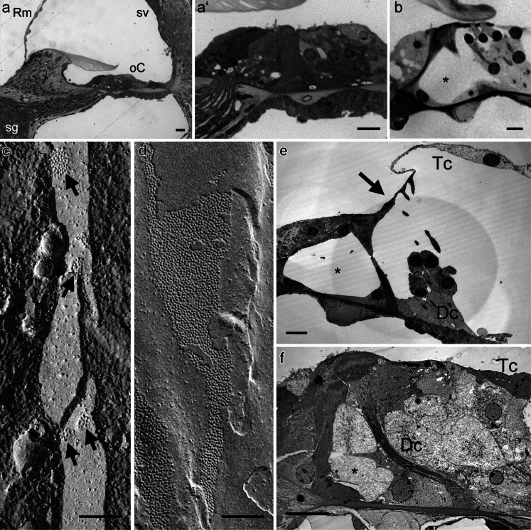
Cx26 and Cx30 play specific roles in development, membrane function and epithelial repair of the cochlea. a, b Light microscopy images of cochlear tissues. a In the basal turn of a 2-week old R75W-Cx26 mouse, the essential sensory and non-sensory tissues are present, but on closer inspection the organ of Corti appears compact (a’). Deiters’ cells are short and intercellular spaces such as the tunnel of Corti and spaces of Nuel are absent. b In comparison, in a juvenile wild-type mouse, the organ of Corti has elongated Deiters’ cells and pillar cells, and a fully patent tunnel of Corti (*). c, d Freeze-fracture images of Deiters’ cell membranes in mice. Freeze-fracture reveals gap junction plaques as clusters of particles in the membrane. Each particle represents an individual channel (connexon). c In Cx30-null animals, plaques of the gap junctions between adjacent Deiters’ cells consist of only a few channels, and they are dispersed. d In wild-type animals, the plaques are extremely large (consisting of thousands of channels) and occupy a significant proportion of the membrane area. e, f Transmission electron micrographs of cochlear tissues. e Following loss of outer hair cells from a Cx30-null mouse, the Deiters’ cells remain columnar in shape, the tunnel of Corti is open (*), and the reticular lamina is displaced towards the pillar cells (arrow). f In a wild-type mouse treated systemically with kanamycin and bumetanide to cause loss of inner and outer hair cells, the Deiters’ cells have expanded to fill the gaps left behind by the hair cells. Cells with characteristics of Deiters’ cells have migrated between the outer pillar cells so that the tunnel of Corti appears filled (*). Dc Deiters’ cell, oC organ of Corti, Rm Reissner’s membrane, sg spiral ganglion, sv stria vascularis, Tc tectal cell. Scale bars (a, b, e, f) 10 μm, (c, d) 0.1 μm

