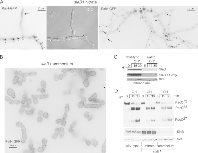
FIG 5.
On ammonium, slaB1 completely prevents PalH endocytosis but does not alter the proteolytic processing activation of PacC. (A) slaB1 hyphae cultured on nitrate, expressing PalH-GFP as described in the legend to Fig. 3. (Left) Hyphae are localized to the plasma membrane at the nascent branch and predominate in the vacuoles (indicated by the letter “v”). (Right) Hyphal tip cells showing weak plasma membrane staining (arrows) and basal conidiospores showing large and conspicuously fluorescent vacuoles. (B) A large field at the same magnification as those used for panel A showing the homogenous population of yeast-like cells resulting from cultivating the slaB1 mutant on ammonium. The PalH-GFP puncta noticeable at the plasma membrane are large pits (not shown). (C) Western blot analysis of SlaB in slaB1 cells cultured on ammonium and shifted from acidic to alkaline pH. The anti-SlaB Western blots in the top and middle panels represent two different exposures (exp) for the same experiment. The lower panel is an anti-hexokinase (hxk) loading control. The middle panel was deliberately overexposed to reveal traces of SlaB. (D) Normal proteolytic processing activation of PacC in slaB1 cells, cultured on ammonium or nitrate, compared to the wild type. As for panel C, the anti-SlaB blot was deliberately overexposed to illustrate the extent of downregulation. Nitrate conditions result in marked overexpression of SlaB.

