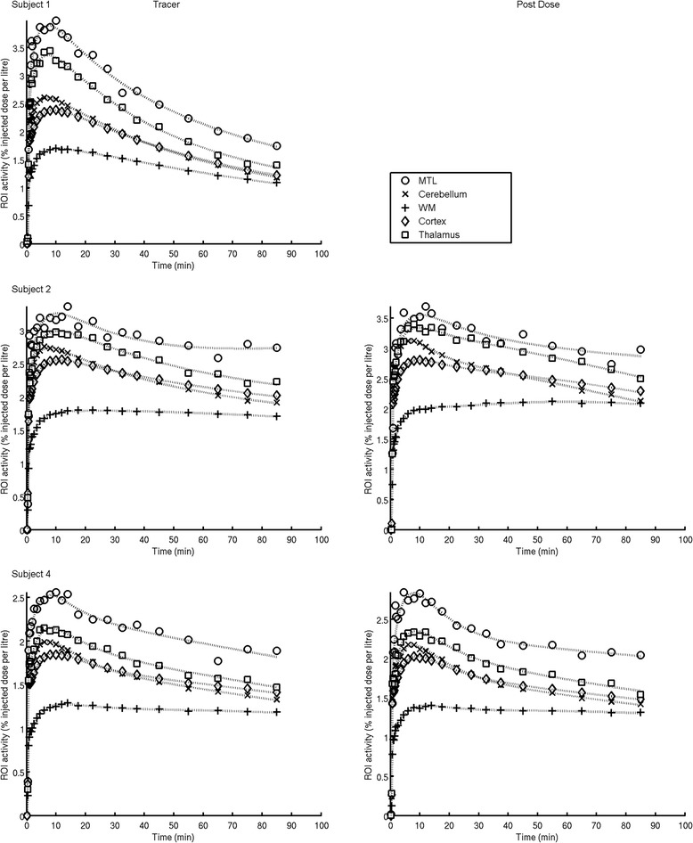
Figure 4.

Regional time activity curves of [ 11 C]GSK1034702 in the human brain and model fits. Regions of interest: white circle, MTL = medial temporal lobe; x, cerebellum; white square, thalamus; diamond, cortex = sum of cortical regions including both cortical grey and white matter; plus sign, WM = all cerebral white matter. Solid lines are two tissue compartmental model fits. For each subject, a regular-sized rectangle (432 mm3) was manually placed over the area of highest signal in the medial temporal lobe (MTL), centred in the hippocampus; this is presented in the data as the MTL ROI.
