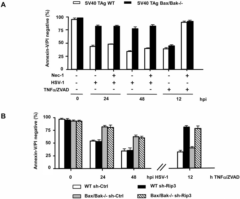
Fig 5. HSV-1-induced cell death does not involve RIP1- and/or RIP3-mediated necroptosis.
(A) Annexin-V/PI FACS analysis of SV40 TAg WT and Bax/Bak-/- MEFs infected with 10 moi of HSV-1 for 0, 24 or 48 h (hpi) or treated with 10 ng/ml TNFα/100μ M ZVAD-fmk ± 100 μM Necrostatin-1 (Nec-1) for 12 h. (B) Annexin-V/PI FACS analysis of mixed populations of SV40 TAg WT and Bax/Bak-/- MEFs stably expressing either sh-Ctrl or sh-Rip3, infected with 10 moi of HSV-1 for 0, 24 or 48 h (hpi) or treated with 10 ng/ml TNFα/100 μM ZVAD-fmk for 12 h. Data are the means of at least three independent experiments ± SEM. The p values are the following: (A) HSV-1-infected Bax/Bak-/- versus WT cells: p < 0.001 for 24 and 48 hpi; TNFα/ZVAD + Nec-1 versus TNFα/ZVAD—Nec-1 for both WT and Bax/Bak-/- cells: p < 0.001; HSV-1 + Nec-1 versus HSV-1—Nec-1 for both WT and Bax/Bak-/- cells: not significant, n = 4. (B) HSV-1-infected Bax/Bak-/- sh-Ctrl versus WT sh-Ctrl and Bax/Bak-/- sh-Rip3 versus WT sh-Rip3: p < 0.001 for 24 and 48 hpi; HSV-1-infected Bax/Bak-/- sh-Ctrl versus Bax/Bak-/- sh-Rip3 and HSV-1-infected WT sh-Ctrl versus WT sh-Rip3: not significant, n = 4. TNFα/ZVAD-treated WT sh-Rip3 versus WT sh-Ctrl and TNFα/ZVAD-treated Bax/Bak-/- sh-Rip3 versus Bax/Bak-/- sh-Ctrl: p < 0.001, n = 3.

