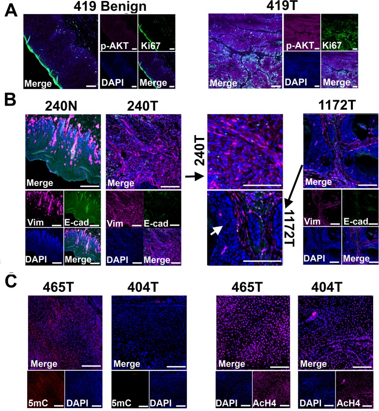
Fig 4. Tissue immunofluorescent staining supports conclusions from RNA-seq studies shown in Fig 3.
(A) Phospho-AKT (p-AKT) is more strongly expressed in tumor cells than in benign cells of canine case 419, indicating the activation of PI3K/AKT pathway in the tumor cells. Tumor 419 has highest AKT1 mRNA expression level based on RNA-seq analysis (Fig 3C). (B) In case 240, tumors cells (240T) express higher level of mesenchymal marker vimentin (Vim) but lower level of epithelial marker E-cadherin (E-cad) when compared to normal squamous cells (240N). Many proliferating tumor cells (e.g., those pointed by white arrow) of case 1172 however do not express vimentin. These are consistent with RNA-seq analyses (Fig 3C). (C) Tumor 404 has significantly decreased 5-methylcytosine (5mC) staining and somewhat decreased acetyl-H4 staining (AcH4), compared to tumor 465. This is consistent with the PCA result shown in Fig 3A indicating them being two distinct tumors. Representative confocal images are shown; scale bar, 50 μm.

