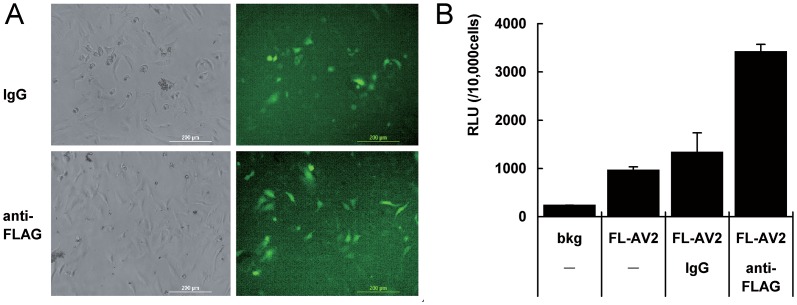
Fig 3. Transduction of immobilized FL-AV2 with a FLAG antibody in 2D HeLa cell cultures.
ELISA plates were pre-coated with bovine type I collagen overnight. Anti-FLAG antibody or non-specific IgG mix was cross-linked collagen by incubation with using activated EDAC and SPDP. The plates were loaded with FL-AV2.GFP overnight and washed 3 times with PBS before use. HeLa cells (5×10⁴ cells/well in 48-well plates) were seeded into the plates and continuingly cultured for 48 h. Fluorescent images were acquired using a Leica DMIRB microscope. (B) Experiments under same conditions of (A) were performed using FL-AV2.Luc viruses for immobilization and infections. The quantitative chemo-luminescence was determined by luciferase assays. Measurements from 6 independent samples were averaged and presented and shown as mean+/-SE.

