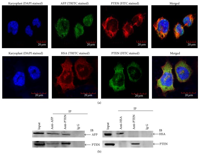
Figure 1.
Colocalization, Coimmunoprecipitation analysis of the interaction of AFP and HSA with PTEN in PLC/PRF/5 cells. (a) Colocalization. Cells were cultured at 37°C in a humidified atmosphere of 5% CO2. Localization of AFP and HSA interaction with PTEN was viewed, and images were captured under laser confocal microscope. Nuclei were stained with DAPI (blue). PTEN were labeled with FITC (green) and AFP and HSA were labeled with FRITC (red), respectively. The image is representative of three independent experiments. (b) Coimmunoprecipitation (Co-IP) analysis of the interaction between AFP and PTEN. Lysates from PLC/PRF/5 cells were immunoprecipitated (IP) with antibodies against HSA, AFP, or PTEN and separated by SDS/PAGE gel electrophoresis. Coimmunoprecipitated complexes were transferred onto a nitrocellulose membrane, immunoblotted with anti-HSA, anti-AFP or anti-PTEN antibody, and scanned by the LAS3000 Chemiluminescence/Fluorescence Instrument (Fuji, Japan). Total protein immunoblotted with antibody against HSA, AFP, or PTEN is defined as input. Images are representatives of an experiment that was repeated three times.

