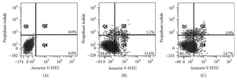
Figure 4.

Flow cytometric analysis of aHSC exposed to SME. aHSCs were exposed to SME at concentrations of 0 (A), 0.3 (B), and 0.5 (C) mg/mL for 72 h. The cells were harvested and incubated with FITC-conjugated annexin V and PI and were measured by flow cytometry. Normal cells were annexin V- and PI-negative (Q3); cells in early apoptosis were annexin V-positive and PI-negative (Q4); cells in late apoptosis/necrosis were annexin V- and PI-positive (Q2).
