. 2015 Jun 3;4:e06359. doi: 10.7554/eLife.06359

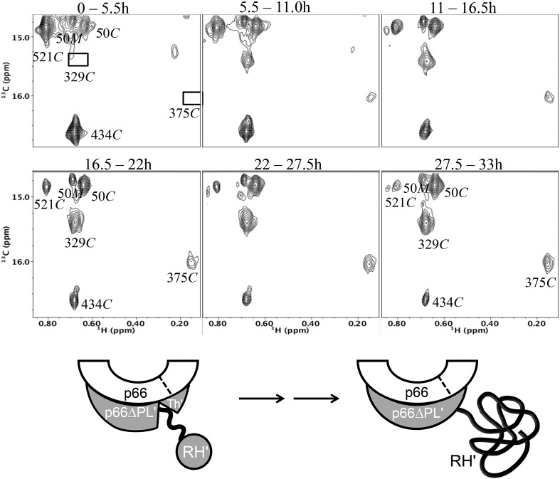
Figure 8. Dimerization of [13CH3-Ile]p66∆PL with unlabeled p66.

Time-dependent changes are shown for a region of the 1H-13C HMQC spectrum covering a similar spectral region to that shown in Figure 7. All resonances are attributed to the M or C species; the labeled Ile50M resonance as well as the Ile434C and 521C resonances arising from the labeled RH' decrease as the RH' domain unfolds, while the connection' 329C and 375C resonances increase as the connection' domain matures. The schematic diagram at the bottom illustrates the subunit-selective labeling pattern and the proposed conformational changes that are inferred from the behavior of the resonances. The labeled subunit is indicated in gray. Each spectrum corresponds to a 5.5-hr accumulation period at the time periods indicated. Dimerization was initiated at t = 0, and the spectra were obtained at 35°C.

DOI: http://dx.doi.org/10.7554/eLife.06359.031

Figure 8.

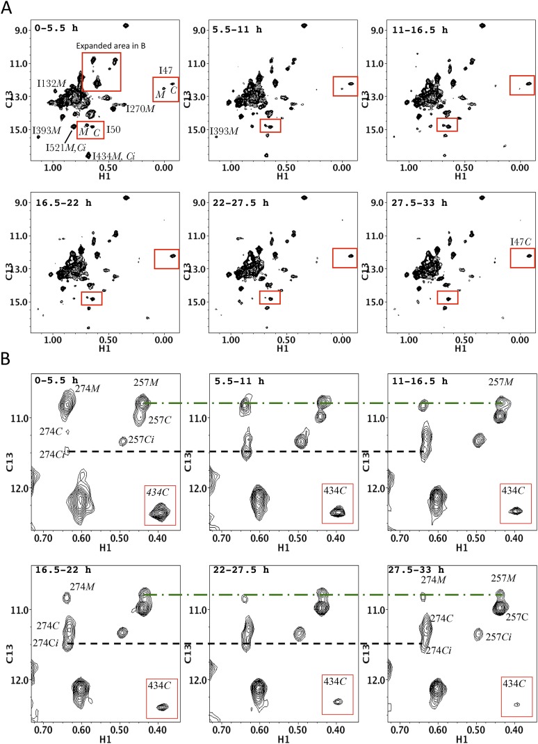
Figure 8—figure supplement 1. Time-dependent HMQC spectra for dimerization of p66∆PL with excess, unlabeled p66 showing all Ile δ-methyl resonances.

Figure 8—figure supplement 1.

(A) Time-dependent spectra for [13CH3-Ile]p66∆PL with a twofold excess of unlabeled p66 after dimerization conditions were initiated. The D2O buffer contained 25 mM Tris-DCl-d11, pD 7.5, 100 mM KCl, 0.02% NaN3. Spectra were obtained on an INOVA 800 at 35 °C. Monomer resonances attributed to Ile50M and to Ile132M that cannot be unequivocally assigned in the homodimerization study can be identified in this study. (B) Expanded region 1 showing the details of the labeled thumb' resonances. The inset illustrates the simultaneous decay of the RH' resonance arising from Ile434.
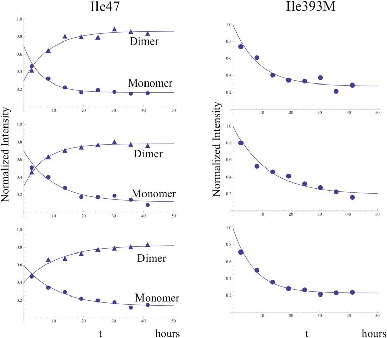
Figure 8—figure supplement 2. Time-dependent intensity data for monomer decay.

Figure 8—figure supplement 2.

Illustrative examples showing the analysis of the time-dependent monomer decay for the Ile47 and Ile393 resonances for the dimerization study illustrated in Figure 8 and two similar studies. In the case of Ile47, decay of the monomer resonance was accompanied by growth of the dimer peak, corresponding to the C conformation, and the sum of the initial Ile47M and Ile47C resonance intensities was normalized to 1.0. The behavior of the Ile47 resonances indicates that an initial, more rapid dimerization process was followed by a slower process with time constant ∼ 9 hr (see Table 2).
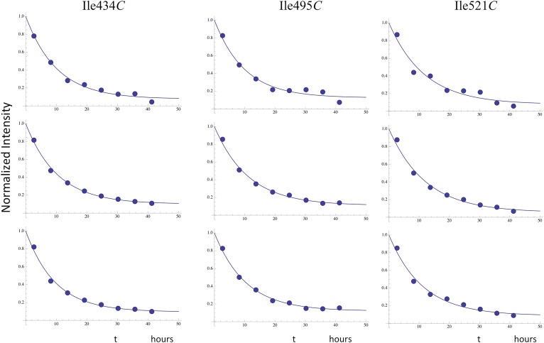
Figure 8—figure supplement 3. Time-dependent decay of RH' resonances.

Figure 8—figure supplement 3.

Illustrative fits of the time-dependent intensity data for the resolved RH' resonances assigned to Ile434C, Ile495C, and Ile521C. The fits allowed for asymptotic monomer resonance intensities greater than 0, and the observed decay generally corresponded to ∼ 90% of the total intensity. The time constants were all in close agreement with a mean value of 10.7 hr (Table 2).
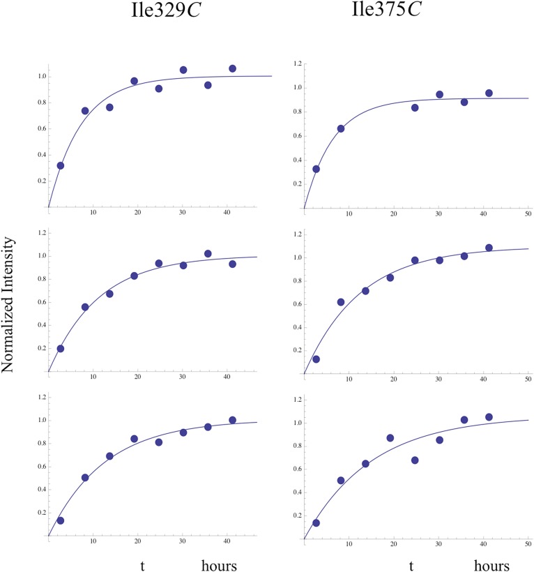
Figure 8—figure supplement 4. Time-dependent growth of connection' resonances.

Figure 8—figure supplement 4.

Fits of the time-dependent intensity data for the resolved Ile329C and Ile375C resonances in the study of the dimerization of [13CH3-Ile]p66∆PL with a twofold excess of unlabeled p66. The Ile375C resonance is significantly weaker than Ile329, so that the sample to sample variation was generally greater.