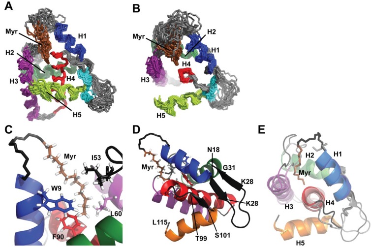
Figure 4.
(A, B) Views of the NMR ensemble (20 structures, generated by superposition of backbone atoms of refined residues) showing the relative orientation of helices and position of the myristyl group; (C, D) Representative NMR structure showing (C) the packing of the myristyl group with the side chains of buried hydrophobic residues; and (D) relative position and orientation of the three-strand pseudo-β-sheet; (E) Comparison of a representative myrMAQ5A/G6S NMR structure with the X-ray crystal structure of myr(-)MA.

