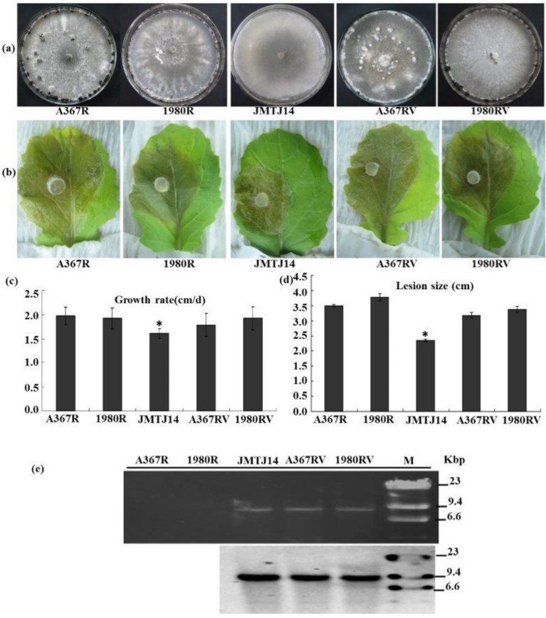
Figure 4.
Biological properties of SsFV1-infected strains of S. sclerotiorum. (a) Colony morphology of S. sclerotiorum strains JMTJ14, A367R (EP-1PNA367R), 1980R, and two new representative SsFV1-infected strains A367RV (EP-1PNA367RV), 1980RV. All S. sclerotiorum strains were grown on PDA for 20 days at 20 °C prior to photography; (b) and (d) Virulence assays of S. sclerotiorum strains listed under detached leaves in (b); Assays were performed on detached rapeseed leaves. The diameters of disease lesion size were measured at 3 days post-inoculation (dpi); (c) Hyphal growth rates of S. sclerotiorum individual strains. Growth rates were examined on PDA at 20 °C; (e) Detection of SsFV1 in individual strains by dsRNA extraction and Northern blot analysis. dsRNA, the replicative form of SsFV1, was extracted and resolved by electrophoresis in a denaturing 1% agarose gel, transferred to a membrane, and probed with specific probe JRP680 (corresponding nt positions 2307–2986) for SsFV1 RNA. The band of about 7.8 kbp in size was clearly detected with dsRNA extraction and Northern blotting. Asterisks indicate a significant difference among strains of S. sclerotiorum according to the Student t test, p = 0.01.

